Introduction
The cell wall is one of the most fundamental structures in biology, a rigid or semi-rigid layer that sits outside the plasma membrane of plant, bacterial, fungal, and algal cells. For every UPSC aspirant working through NCERT biology, understanding the cell wall is essential because it links directly to topics in plant physiology, microbiology, antibiotics, food production, and even biotechnology questions that routinely appear in Prelims and GS Paper III.
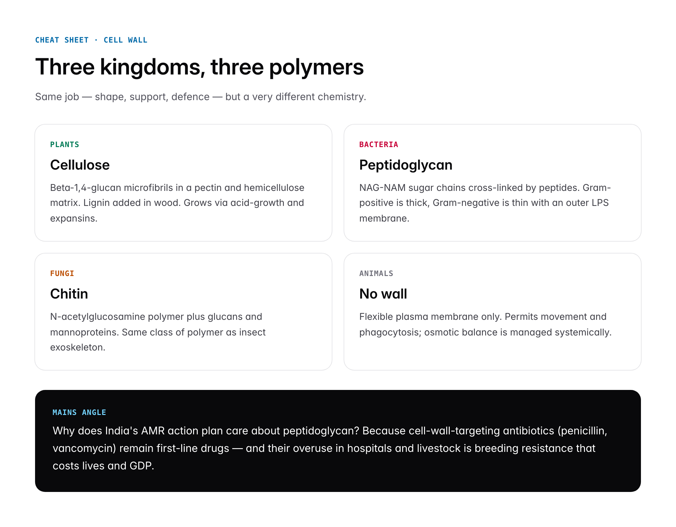
Unlike the delicate plasma membrane, the cell wall gives a cell its shape, protects it from bursting under osmotic pressure, and mediates how a cell interacts with its neighbours. Yet the composition of this wall is not uniform across living organisms. Plants build it from cellulose, bacteria from peptidoglycan, and most fungi from chitin. These differences are not trivia; they underpin crop science, targeted antibiotics, and the entire classification of microbial life.
Quick Facts at a Glance
| Attribute | Detail |
|---|---|
| Found in | Plants, bacteria, fungi, algae, some archaea |
| Absent in | Animal cells, mycoplasma, protozoa |
| Plant main polymer | Cellulose (beta-1,4-glucan) |
| Bacterial main polymer | Peptidoglycan (murein) |
| Fungal main polymer | Chitin |
| Discovery | Robert Hooke, 1665, observed in cork |
| NCERT coverage | Class 9 Ch. 5; Class 11 Biology Ch. 8 |
| Key UPSC link | Antibiotics (penicillin), Gram staining, bio-fortification |
Background and Historical Context
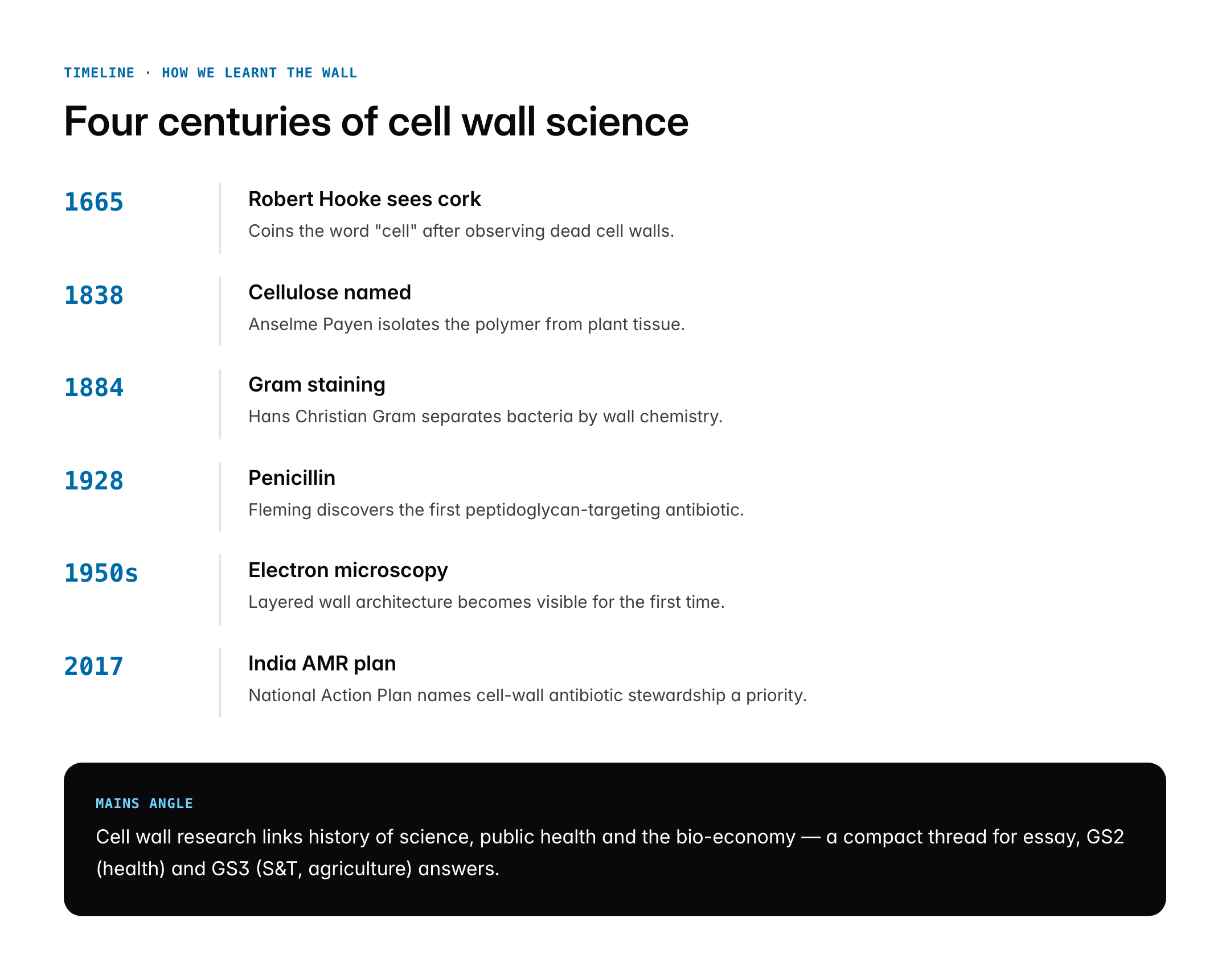
The cell wall was actually the first cellular structure ever seen. In 1665, Robert Hooke examined a thin slice of cork under his primitive microscope and observed tiny empty chambers that reminded him of monks’ cells in a monastery. What Hooke saw were in fact the dead, lignified cell walls of cork tissue, the living contents having long since dried out. The word “cell” therefore entered biology not to describe cytoplasm but to describe the wall that enclosed it.
For nearly two centuries the cell wall was treated as a passive container. In the 19th century, as Matthias Schleiden and Theodor Schwann formulated the Cell Theory, botanists began to notice that plant cells had walls of distinct chemical character, rich in what Anselme Payen in 1838 isolated and named cellulose. Bacteriologists in the late 19th century, led by Hans Christian Gram in 1884, discovered that bacteria could be separated into two great groups based on how their walls retained a violet dye. This simple staining method, still used every day in pathology labs, turned the cell wall into a clinical tool.
The 20th century brought molecular detail. Alexander Fleming’s discovery of penicillin in 1928 and later structural work by Dorothy Hodgkin exposed how the drug attacked bacterial peptidoglycan, sparing human cells because animals have no wall at all. Electron microscopy from the 1950s onwards revealed layered architecture in plants, middle lamella, primary wall, secondary wall, and showed that walls are dynamic structures that grow, thicken, and can be remodelled in response to hormones, pathogens and environmental stress. Today the cell wall is an active field of research in bio-energy, where cellulose and hemicellulose are broken down to produce second-generation biofuels.
Key Features
The cell wall looks simple under a light microscope, but at molecular resolution it is a sophisticated composite material, comparable in engineering terms to reinforced concrete.
Plant Cell Wall
The plant cell wall is built around long, unbranched chains of cellulose, a polymer of thousands of glucose units linked by beta-1,4 glycosidic bonds. These chains bundle into microfibrils, which are embedded in a matrix of hemicellulose, pectin, and in older cells, lignin. There are three recognised layers: the middle lamella shared between adjacent cells and rich in pectin; the primary wall laid down during growth; and the secondary wall, deposited inside the primary wall once growth stops, often strengthened with lignin to form wood.
Bacterial Cell Wall
The bacterial cell wall is chemically different and diagnostically vital. Its principal molecule is peptidoglycan or murein, a mesh of sugar chains (N-acetylglucosamine and N-acetylmuramic acid) cross-linked by short peptides. Gram-positive bacteria such as Staphylococcus carry a thick peptidoglycan layer of 20 to 80 nanometres, while Gram-negative bacteria such as E. coli have a thin peptidoglycan layer (5 to 10 nm) wrapped in an additional outer membrane containing lipopolysaccharide. This outer membrane is the reason Gram-negative infections are often harder to treat.
Fungal Cell Wall
The fungal cell wall uses a third polymer, chitin, the same tough nitrogen-containing polysaccharide that makes insect exoskeletons. Fungi also incorporate glucans and mannoproteins. This chitin-rich wall is a key target for antifungal drugs such as echinocandins.
Archaeal and Algal Walls
Archaea often lack peptidoglycan and use pseudopeptidoglycan or S-layer proteins, which is one reason many antibiotics that target bacteria do not affect them. Algae vary widely, diatoms use silica, red algae use agar and carrageenan, and brown algae use alginate, all of which have major industrial value.
Significance for UPSC and General Knowledge
- The cell wall is a recurring Prelims topic, especially its composition across kingdoms, a classic one-mark MCQ.
- It is the structural basis of the Gram staining technique that forms the foundation of clinical microbiology and antibiotic selection.
- Penicillin, cephalosporins and vancomycin all act by disrupting peptidoglycan synthesis, a key example for GS Paper III questions on science and public health.
- Cellulose is the most abundant organic polymer on Earth, central to textiles (cotton is over 90 percent cellulose), paper, and second-generation biofuels.
- Lignin in secondary walls is the reason wood, a renewable resource, has mechanical strength, and lignin degradation is a frontier in bioethanol research.
- Cell wall biology links directly to Indian missions in agriculture (bio-fortification), bio-economy (ethanol blending), and public health (antimicrobial resistance).
Detailed Analysis: Functions and Biological Roles
The cell wall performs at least six distinct functions that together make multicellular and microbial life possible. First and most obvious is mechanical support. A plant cell in full turgor can experience internal pressures of five to ten atmospheres, comparable to a car tyre, and only the wall prevents the membrane from bursting. In woody plants, lignified secondary walls enable trees to stand tens of metres tall against gravity and wind. Second is protection. The wall is the first line of defence against pathogens, herbivores and physical damage, and it releases signalling fragments called oligosaccharins when attacked, triggering the plant’s immune response.
Third is shape determination. The orientation of cellulose microfibrils, controlled by cortical microtubules inside the cell, dictates whether a cell elongates, expands radially, or remains cuboidal. This is how a root tip cell knows to become a long epidermal cell rather than a spherical storage cell. Fourth is transport and communication. Plant walls are perforated by plasmodesmata, membrane-lined channels that connect neighbouring cytoplasms and allow water, ions, and even RNA molecules to flow between cells in a continuous symplast.
Fifth, the wall regulates water and solute movement. The apoplast, the interconnected network of cell walls and intercellular spaces, is a major pathway for water transport in roots until blocked by the Casparian strip in the endodermis. Sixth, in bacteria, the wall is an identity marker, carrying surface antigens that the human immune system recognises and that vaccines target; the pneumococcal vaccine works by training the immune system against wall polysaccharides. In fungi, similar wall molecules drive both allergic responses and therapeutic targets for antifungals.
Beyond these classical roles, recent research has shown that cell walls are active in sensing. Plants can detect touch, wounding, and even drought through wall integrity pathways, and many crop traits from lodging resistance in wheat to fibre quality in cotton map back to wall biology.
Comparative Perspective
The differences between plant, bacterial and fungal walls are frequently asked in UPSC Prelims and also appear in competitive exams like CSIR-NET and NEET. A side-by-side comparison clarifies the picture.
| Feature | Plant | Bacteria | Fungi |
|---|---|---|---|
| Main polymer | Cellulose | Peptidoglycan | Chitin |
| Secondary components | Hemicellulose, pectin, lignin | Teichoic acids, LPS | Glucans, mannoproteins |
| Typical thickness | 0.1 to 10 micrometres | 10 to 80 nanometres | 100 to 200 nanometres |
| Permeability | High to water, selective to solutes | Variable, LPS in Gram-negative | Moderate |
| Drug target | Herbicides (cellulose synthase inhibitors) | Antibiotics (penicillin) | Antifungals (echinocandins) |
| Growth mechanism | Acid growth via expansins | Autolysins remodel peptidoglycan | Tip growth with chitin synthases |
Animal cells, by contrast, have no wall, which gives them flexibility for movement and phagocytosis but makes them vulnerable to osmotic lysis, a limitation compensated by circulatory regulation of body fluids.
Controversies and Debates
Cell wall biology is not free of scientific controversy. The first long-running debate concerns wall growth. The classical acid-growth hypothesis, proposed in the 1970s, holds that the plant hormone auxin triggers proton pumping into the wall, acidifying it and activating expansins that loosen cellulose cross-links. Critics argue the mechanism is oversimplified and that multiple wall sensors and feedback loops operate in parallel.
A second debate surrounds antimicrobial resistance. Overuse of cell-wall-targeting antibiotics, especially in Indian hospitals and livestock, has selected for resistant strains like methicillin-resistant Staphylococcus aureus (MRSA) and vancomycin-resistant Enterococci. The World Health Organization lists AMR among the top ten public health threats, and India’s National Action Plan on AMR (2017) identified cell wall antibiotic stewardship as a priority.
A third controversy is genetically modified crops with altered cell walls. Reduced-lignin plants are easier to convert into bioethanol, but critics fear weaker stems and higher pest susceptibility. Bt cotton and similar biotech crops raise related public policy debates in India that recur in GS Paper III. Finally, the commercial push to patent cellulose nanofibres and nanocellulose composites has triggered concern about biopiracy and technology access in developing countries.
Prelims Pointers
- The term cell was coined by Robert Hooke in 1665 while observing cork cell walls.
- Cellulose is a homopolymer of beta-D-glucose linked by beta-1,4 glycosidic bonds.
- Middle lamella is rich in calcium and magnesium pectates.
- Gram-positive bacteria retain crystal violet because of their thick peptidoglycan.
- Lysozyme, an enzyme in tears and saliva, breaks peptidoglycan and is a natural antibiotic.
- Penicillin targets the enzyme transpeptidase involved in peptidoglycan cross-linking.
- Mycoplasma has no cell wall and is resistant to penicillin.
- Chitin is a polymer of N-acetylglucosamine and is present in fungi and arthropods.
- Diatoms have silica-based frustules instead of polysaccharide walls.
- Plasmodesmata are analogous to gap junctions in animal cells.
- Suberin and cutin are hydrophobic materials deposited in specialised plant walls.
- The primary cell wall is laid down during cell division; the secondary wall after growth stops.
Mains Practice Questions
- Discuss the structural and functional differences between plant, bacterial and fungal cell walls, and explain how these differences are exploited in modern medicine and agriculture. (250 words)
- Compare composition: cellulose, peptidoglycan, chitin; layered architecture in plants; Gram classification in bacteria.
- Link to antibiotics (penicillin, vancomycin), antifungals (echinocandins), herbicides targeting cellulose synthase.
- Bring in AMR, bio-fortification, and second-generation biofuels as Indian policy angles.
- “Understanding the cell wall is central to both public health and the bio-economy.” Examine this statement with Indian examples. (250 words)
- Cell-wall antibiotics and India’s National Action Plan on AMR.
- Cellulose in cotton textiles, paper industry, and ethanol blending targets.
- Emerging areas: nanocellulose, agricultural residue-based biofuels, fungal biotech.
Conclusion
The cell wall is far more than a passive shell. It is a living composite of cellulose, peptidoglycan or chitin that gives cells their shape, protects them from pathogens, enables communication between neighbours, and sets many of the boundaries of life on Earth. From the monastic cells that Robert Hooke sketched in 1665 to today’s laboratories synthesising nanocellulose for electronics, the wall has remained a central object of scientific curiosity.
For an aspirant, the cell wall sits at the intersection of basic biology and contemporary policy. It explains why penicillin saves lives and why its misuse breeds resistance; why cotton dominates Indian textiles; why lignin slows the biofuel revolution; and why fungi deserve dedicated antifungal drug pipelines. A strong grasp of cell wall structure therefore pays dividends in both Prelims MCQs and Mains essays on science, technology, and the Indian bio-economy.
Frequently Asked Questions
What is a cell wall?
A cell wall is a rigid or semi-rigid layer that surrounds the plasma membrane of plant, bacterial, fungal, and algal cells. It is built from polymers such as cellulose in plants, peptidoglycan in bacteria, and chitin in fungi. The wall provides shape, mechanical strength, osmotic protection, and a first line of defence against pathogens and environmental stress.
Why is the cell wall important for UPSC?
The cell wall appears in Prelims questions on cell biology and microbiology, in Gram staining, and in Mains topics covering antibiotics, antimicrobial resistance, bio-fortification and biofuels. Understanding its composition across kingdoms links to India’s National Action Plan on AMR and to agricultural and bio-economy policy questions in GS Paper III.
How is the cell wall related to antibiotics like penicillin?
Penicillin and related beta-lactam antibiotics target the enzyme transpeptidase that cross-links peptidoglycan, the main polymer of bacterial cell walls. Because human cells lack a wall, the drug kills bacteria without harming patients. Excessive use, however, selects for resistance, which is why cell wall biology is central to antimicrobial stewardship.
What is the main component of a plant cell wall?
Cellulose is the main component of the plant cell wall. It is a linear polymer of beta-D-glucose units linked by beta-1,4 glycosidic bonds, bundled into microfibrils. These microfibrils are embedded in a matrix of hemicellulose, pectin, and in mature cells, lignin, forming a composite that rivals engineered materials in tensile strength.
How do Gram-positive and Gram-negative bacteria differ in their walls?
Gram-positive bacteria have a thick peptidoglycan layer (20 to 80 nm) that retains crystal violet dye during Gram staining, appearing purple. Gram-negative bacteria have a thin peptidoglycan layer (5 to 10 nm) plus an outer membrane containing lipopolysaccharide, stain pink, and are often more resistant to antibiotics because of this extra barrier.
Do animal cells have a cell wall?
No, animal cells do not have a cell wall. They are bounded only by a flexible plasma membrane, which allows shape change, movement, and phagocytosis. The absence of a wall is compensated by circulatory regulation of body fluids to prevent osmotic lysis and is one reason many cell-wall-targeting antibiotics are safe for humans.
What is plasmodesmata?
Plasmodesmata are narrow, membrane-lined channels that traverse the walls between neighbouring plant cells. They connect the cytoplasms of adjacent cells into a continuous network called the symplast, allowing water, ions, small molecules and even regulatory RNAs to move directly between cells. They are functionally analogous to gap junctions in animal tissues.
Why does mycoplasma not respond to penicillin?
Mycoplasma is a genus of bacteria that naturally lacks a cell wall. Because penicillin kills bacteria by disrupting peptidoglycan synthesis, and mycoplasma has no peptidoglycan to disrupt, the drug has no target. Mycoplasma infections such as atypical pneumonia are therefore treated with antibiotics like macrolides or tetracyclines that attack protein synthesis instead.