

Why in News?
On 15 October 2026, the Technology Development Board announced an agreement with Panacea Medical Technologies to support development and commercialisation of an indigenous 32-slice computed tomography scanner.
- TDB, under the Department of Science and Technology, is supporting the Karnataka-based manufacturer’s diagnostic imaging project.
- The announcement concerns development and commercialisation; it does not establish completed deployment, clinical validation or regulatory clearance.
- The project combines precision engineering, imaging components and calibration; no verified final selling price or patient-dose advantage was announced.
- Domestic capability in medical equipment can reduce dependence on imported technologies, but manufacturing origin alone cannot establish diagnostic quality.
- For public healthcare, access requires usable equipment, trained teams and reliable maintenance, alongside appropriate decisions about when imaging is necessary.
UPSC Relevance
Prelims Relevance
- Computed tomography and cross-sectional imaging
- X-rays as ionising radiation
- Digital detectors and image reconstruction
- CT versus MRI
- TDB under the Department of Science and Technology
Mains Relevance
GS Paper 3
- Indigenous medical technology and quality assurance
- Engineering capability beyond final assembly
GS Paper 2
- Diagnostic access and patient safety
Essay
- Useful innovation must combine wider access with trustworthy performance.
Background and Context
What the Indigenous Scanner Project Establishes
The agreement creates a development opportunity; the clinically useful product must still be distinguished from the announcement that supports its creation.
- The PIB announcement identifies an indigenous 32-slice CT scanner as the project objective. It describes planned design, development and deployment, rather than demonstrating that a finished scanner is already serving patients.
- Commercialisation means taking technology towards practical market use. Support for this stage is different from evidence that the equipment has obtained every necessary permission or demonstrated safety and effectiveness in clinical operation.
- Indigenous capability involves integrating imaging components with engineering and software. The relevant policy question is whether domestic expertise can sustain reliable equipment, rather than merely whether a machine carries an Indian manufacturer’s name.
- The 32-slice label identifies the announced scanner category; it should not be read as a complete statement of image resolution. Assessment needs actual performance evidence, which the announcement does not supply.
- Access is the intended public benefit, not an already measured result. Lower import dependence could strengthen equipment availability, but claims of cheaper scans or better outcomes require evidence beyond the signed development agreement.
How X-Ray Measurements Become Cross-Sectional Images
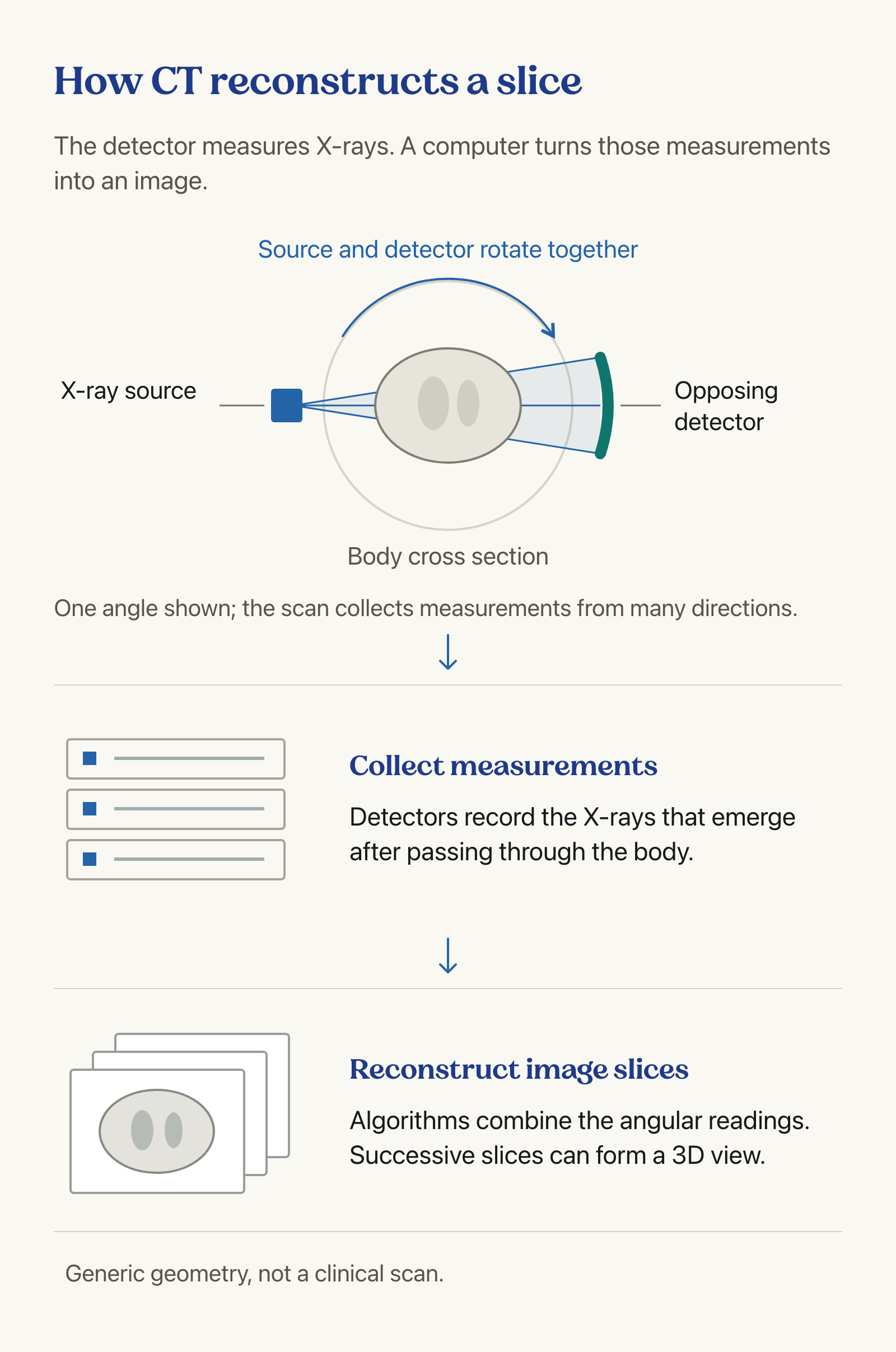
Computed tomography measures how X-rays pass through the body from different angles, then uses computation to reconstruct internal structures.
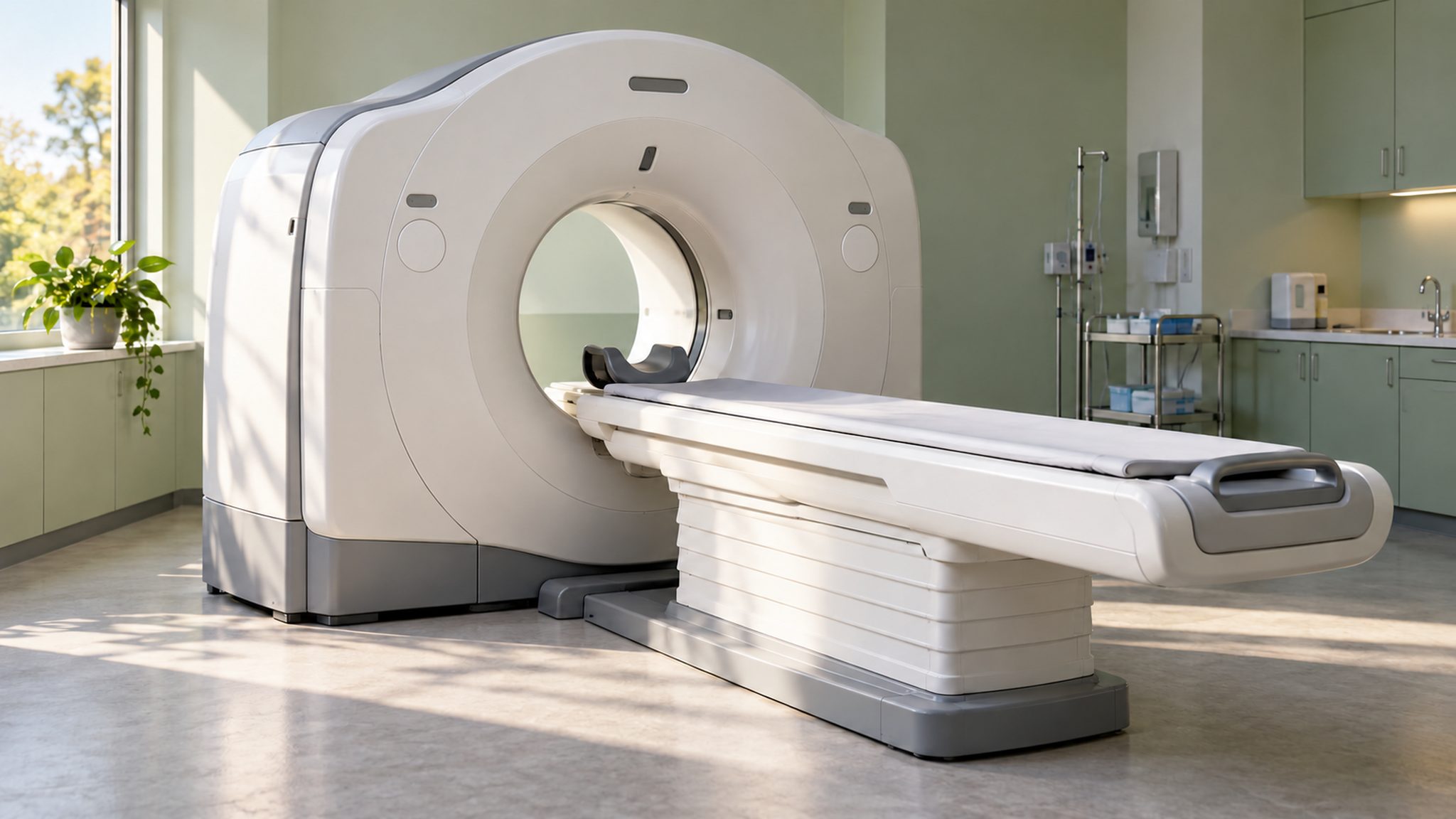
- A rotating X-ray source sends beams through the body while the patient lies on a moving table. The circular gantry houses the imaging system; the external opening is not itself the detector.
- Digital detectors register X-rays emerging from the body. Measurements collected at different angles provide information about internal structures; a single detector reading is not yet the cross-sectional picture that a clinician interprets.
- Reconstruction algorithms combine the angular measurements into image slices. As the NIBIB explanation describes, these slices can also be stacked into three-dimensional views, making computation central to the imaging process.
- Calibration connects precise engineering to trustworthy measurement. The development announcement specifically identifies rigorous calibration as necessary for imaging accuracy and patient safety; producing an image alone does not establish that the system performs reliably.
- Diagnostic interpretation follows reconstruction: the scanner supplies images for clinical assessment, rather than an automatic verdict about disease. This distinction matters when discussing technology-led healthcare, where useful information still requires professional judgment and appropriate follow-up.
Radiation Safety and the CT–MRI Distinction
The objective is an examination that answers a clinical question with appropriate exposure, rather than either avoiding all imaging or maximizing scanning.
- CT uses ionising radiation, which can increase lifetime cancer risk. The FDA explains that benefits generally outweigh risks when the examination is medically necessary; risk awareness must not become blanket rejection of useful diagnosis.
- Justification asks whether CT is needed to answer the clinical question. Reviewing earlier imaging and considering suitable alternatives can prevent unnecessary repeat examinations without treating every request for diagnostic information as equally urgent.
- Optimization means selecting exposure that produces an image adequate for the diagnostic task. The lowest possible exposure is not automatically the best setting if the resulting image cannot answer the clinician’s question.
- Children need appropriate exposure settings because their radiation risk differs from adults. Safety depends on the imaging team and examination protocol as well as the scanner’s hardware, making training part of responsible deployment.
- MRI does not use ionising radiation, but this does not mean it is risk-free or interchangeable with CT. FDA guidance identifies implant-related hazards; modality choice must follow the clinical question and patient circumstances.
Way Forward
Evaluate Performance Before Scaling Access
- Require documented performance and safety evidence before describing development support as a completed diagnostic breakthrough.
- Build calibration and maintenance capability alongside manufacturing so hospitals can sustain reliable equipment throughout its working life.
- Train imaging teams in justification and exposure optimization, including appropriate protocols for children and review of previous examinations.
Conclusion
- CT combines physical measurement with mathematical reconstruction: rotating X-rays produce detector data, and computation converts that information into slices. Neither a development agreement nor a slice-count label substitutes for demonstrated diagnostic performance.
- For healthcare policy, judge self-reliance by dependable equipment and useful access together. Manufacturing support becomes valuable when reliable imaging reaches patients through trained teams, appropriate examinations and sustained quality assurance.
UPSC Practice Questions
Prelims MCQ 1
With reference to computed tomography, consider the following statements:
- It uses ionising radiation.
- Its computers reconstruct cross-sectional images from measurements obtained at different angles.
- MRI and CT use the same physical imaging principle.
How many of the above statements are correct?
(a) Only one (b) Only two (c) All three (d) None
Answer: (b) Only two
Explanation:
CT uses X-rays and computational reconstruction. MRI does not use ionising radiation and has a different physical imaging principle.
Prelims MCQ 2
Which statement best describes exposure optimization in CT imaging?
(a) Using maximum exposure for every patient (b) Avoiding every medically necessary examination (c) Using exposure adequate for the diagnostic task while minimizing unnecessary radiation (d) Replacing clinical interpretation with slice count
Answer: (c) Using exposure adequate for the diagnostic task while minimizing unnecessary radiation
Explanation:
Optimization balances radiation exposure with sufficient image quality; justification separately addresses whether the examination is needed.
UPSC Mains Questions
- Explain how computed tomography converts X-ray measurements into images. Discuss the role of calibration and exposure optimization in patient safety.
- Indigenous medical-device manufacturing must be assessed beyond import substitution. Discuss with reference to diagnostic imaging equipment.
Sources: PIB, Ministry of Science and Technology and NIBIB, National Institutes of Health.
Frequently Asked Questions
What has TDB announced about the indigenous CT scanner?
TDB has announced support for development and commercialisation of a 32-slice CT scanner by Panacea Medical Technologies. This is not evidence of completed deployment, clinical validation or regulatory clearance.
How does a CT scanner create slices?
An X-ray source rotates around the body, and digital detectors collect measurements from multiple angles. A computer reconstructs these measurements into cross-sectional images that can be viewed individually or combined.
Does a 32-slice label prove higher image resolution?
No. The label alone does not provide a complete measure of diagnostic performance or resolution. The announced project needs performance evidence; the release does not establish numerical resolution or superiority.
How does CT differ from MRI?
CT uses X-rays and ionising radiation. MRI does not use ionising radiation, but has other safety considerations, including hazards involving some implants. They are not automatically interchangeable for every clinical question.
What does CT exposure optimization mean?
It means selecting exposure that provides image quality adequate for the diagnostic task while avoiding unnecessary radiation. Appropriate settings, trained teams and attention to patient characteristics are central to this approach.
Tell Google you want more of this.
Add Anantam IAS as a preferred sourceOne tap, and this site shows up more often in your own Top Stories, AI Overviews and AI Mode. Remove it any time.