The immune system is, at its core, a standing army that has been handed live ammunition and told to distinguish friend from foe at the level of individual molecules. It does this remarkably well most of the time. But the same machinery that destroys a virus-infected cell can, if uncontrolled, turn on the pancreas and cause type 1 diabetes, or attack the joints and produce rheumatoid arthritis, or strip the protective sheath from nerves and lead to multiple sclerosis. The puzzle of how the body keeps this army on a leash, attacking pathogens but sparing its own tissues, is one of the deepest in immunology. The answer, worked out over the last three decades, has a name: peripheral immune tolerance, enforced by a small but powerful cell population called regulatory T cells, or Tregs.
The master switch that turns an ordinary T cell into a Treg is a single gene, FOXP3, sitting on the X chromosome. Without a functional FOXP3, Tregs cannot form, and the immune system loses its brakes. With too many Tregs in the wrong place, such as inside a tumour, the immune system loses its will to attack. The biology of FOXP3 and Tregs is, in this way, the biology of restraint, and modern medicine, from cancer immunotherapy to transplant tolerance to the treatment of fatal infant autoimmunity, increasingly turns on understanding it.
For UPSC, FOXP3 and Tregs sit at the intersection of GS-III science and technology, contemporary biology questions, and current affairs anchored to the 2025 Nobel Prize in Physiology or Medicine, which honoured the discovery of peripheral immune tolerance and the role of regulatory T cells.
Quick Facts: FOXP3 and Tregs at a Glance
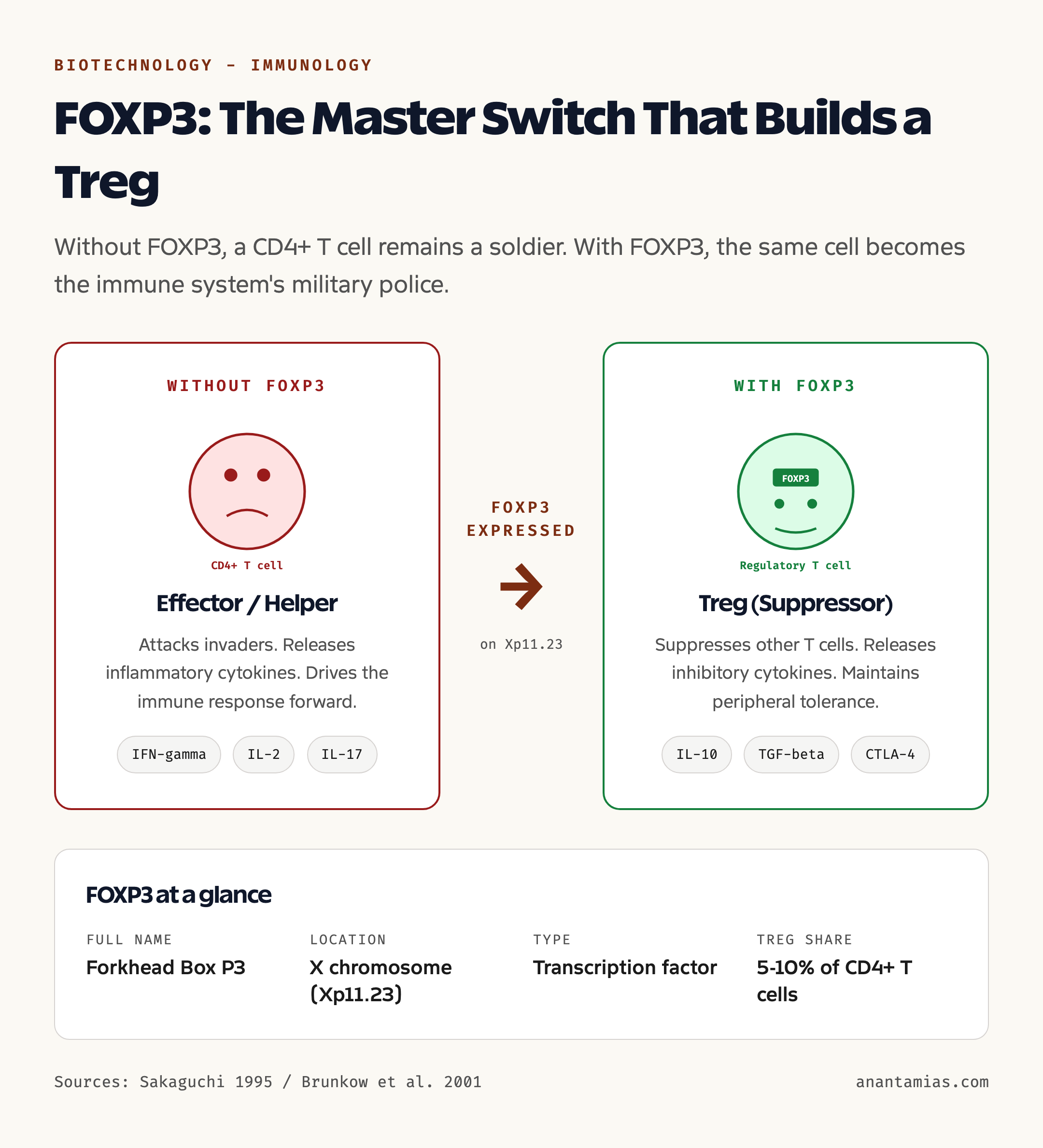
- FOXP3 full name: Forkhead Box P3
- Gene location: X chromosome (Xp11.23)
- Function: Master transcription factor for regulatory T cells
- Cell type controlled: CD4+ regulatory T cells (Tregs)
- Treg share of CD4+ T cells: Roughly 5 to 10 percent in healthy adults
- Key role: Maintain peripheral immune tolerance and prevent autoimmunity
- Loss-of-function disease: IPEX syndrome (Immunodysregulation Polyendocrinopathy Enteropathy X-linked)
- Discovery of Tregs: Shimon Sakaguchi, 1995
- FOXP3 link to Tregs: Established in the early 2000s, including work by Mary Brunkow and Fred Ramsdell on the scurfy mouse
- Recognition: 2025 Nobel Prize in Physiology or Medicine for peripheral immune tolerance and regulatory T cells, awarded as per latest available data to Mary Brunkow, Fred Ramsdell and Shimon Sakaguchi
- Cancer connection: Tumours recruit Tregs to suppress anti-tumour immunity
- Therapeutic frontier: Treg-depleting and Treg-expanding therapies in autoimmunity, transplantation and oncology
What Are Tregs and What Does FOXP3 Do?
A T cell starts life in the bone marrow, matures in the thymus and circulates through the blood and lymph nodes searching for trouble. Most T cells are effectors. The CD8+ killer T cells destroy infected or malignant cells directly. The CD4+ helper T cells coordinate the rest of the immune response, calling in B cells to make antibodies and macrophages to mop up debris. These are the soldiers of the immune army.
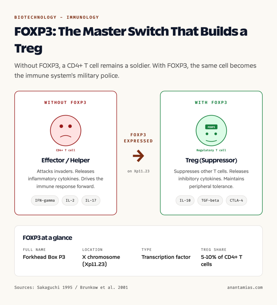
Regulatory T cells are a different kind of T cell. They are CD4+, like helpers, but they carry an additional surface marker called CD25 and, crucially, express the FOXP3 gene at high levels. Their job is not to attack, but to suppress. They release inhibitory cytokines such as IL-10 and TGF-beta, they consume IL-2 that other T cells need to grow, and they make direct contact with effector T cells to switch them off. The metaphor that has become standard is that of military police: the soldiers go out to fight, the police make sure they stop fighting once the threat is gone and that they never turn their weapons on the citizens, in this case the body’s own organs.
FOXP3 is the badge that confers police authority. It is a transcription factor, which means it is a protein that binds to DNA and turns thousands of other genes on or off. When a developing T cell expresses FOXP3 at sufficient levels, it commits to the regulatory lineage, takes on suppressive function, and acquires the ability to enforce tolerance. Without FOXP3, a T cell remains an effector. The fate of the entire Treg population, and through it the integrity of peripheral immune tolerance, hinges on this one gene.
Background and Historical Context
The intuition that the immune system needs a brake is older than its molecular proof. Through the 1970s and 1980s, immunologists noticed that animals depleted of certain T cell subsets developed autoimmune disease, and that tolerance to self could be transferred from one animal to another by transferring T cells. The cells responsible were elusive. Various candidates, including a poorly defined population called suppressor T cells, came and went without surviving rigorous reanalysis. By the late 1980s, the field was sceptical that a dedicated suppressor lineage existed at all.
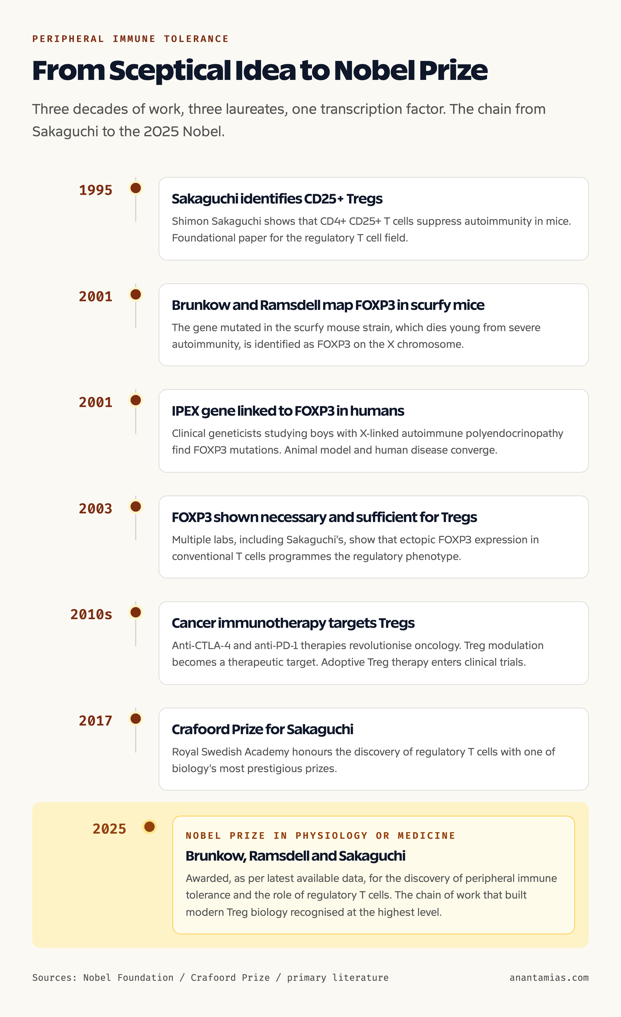
Shimon Sakaguchi changed that in 1995. Working at Nagoya University, he and his colleagues showed that CD4+ T cells expressing the IL-2 receptor alpha chain, CD25, formed a distinct population that could prevent autoimmune disease when transferred into immunodeficient hosts. Removing this population from healthy mice caused them to develop multi-organ autoimmunity. The brake, in other words, was real, and it was a defined cell type. This was the foundational discovery of regulatory T cells.
The molecular basis remained unclear for almost a decade. Two parallel lines of work cracked it. Mary Brunkow, Fred Ramsdell and colleagues studying the scurfy mouse, a mutant strain that died young from severe autoimmunity, mapped the responsible gene to FOXP3 in 2001. At the same time, clinical geneticists studying boys with IPEX syndrome, an X-linked disease causing fatal infant autoimmunity, found the same gene mutated. By 2003, several laboratories, including Sakaguchi’s, had shown that FOXP3 was both necessary and sufficient to programme the regulatory phenotype. A T cell that expressed FOXP3 became a Treg; one that did not could not. The connection between Sakaguchi’s cell and the Brunkow-Ramsdell gene closed the loop.
The recognition came in stages. The 2017 Crafoord Prize honoured Sakaguchi for the discovery of Tregs. The 2025 Nobel Prize in Physiology or Medicine, as per latest available data, was awarded for the discovery of peripheral immune tolerance and the role of regulatory T cells, with Mary Brunkow, Fred Ramsdell and Shimon Sakaguchi recognised for the chain of work that took the field from a contested idea to a foundation of modern medicine. For background on the scientific infrastructure that supports this kind of work in India, see our coverage of biotechnology and the pre-clinical research network.
Key Mechanism: How FOXP3 Builds a Treg
Tregs come in two main types. Thymic Tregs, sometimes called natural Tregs, are programmed during T cell development in the thymus. As thymocytes mature and encounter self-antigens displayed by thymic epithelial cells, those whose T cell receptors bind self with intermediate strength are diverted into the Treg lineage. FOXP3 is induced during this process and locks in the regulatory fate. These thymic Tregs leave the thymus already pre-committed to suppressing autoreactive responses in the periphery.
Peripheral or induced Tregs arise outside the thymus, in lymph nodes and tissues, when conventional CD4+ T cells encounter antigen in the presence of TGF-beta and other tolerogenic signals. Under those conditions, FOXP3 is switched on and the cell converts to a Treg. This pathway is particularly important at mucosal surfaces, where the gut and respiratory tract are constantly exposed to harmless food proteins and commensal bacteria. Without induced Tregs, the immune system would react to every meal as an infection.
Once FOXP3 is expressed, it works as part of a transcriptional complex with other regulators to switch on a suppressive programme. Treg-defining genes such as CD25, CTLA-4, and the inhibitory cytokines IL-10 and TGF-beta come on. Effector T cell genes are repressed. The cell adopts a metabolism distinct from that of conventional T cells, relying more on oxidative phosphorylation than on glycolysis. The combined effect is a cell that, on encountering an antigen, dampens the response of nearby T cells rather than amplifying it. Stable FOXP3 expression depends on the demethylation of a specific region in the FOXP3 locus called the Treg-specific demethylated region, an epigenetic signature that distinguishes a committed Treg from a transiently FOXP3-expressing conventional cell.
Why It Matters: Tolerance, Autoimmunity and Cancer
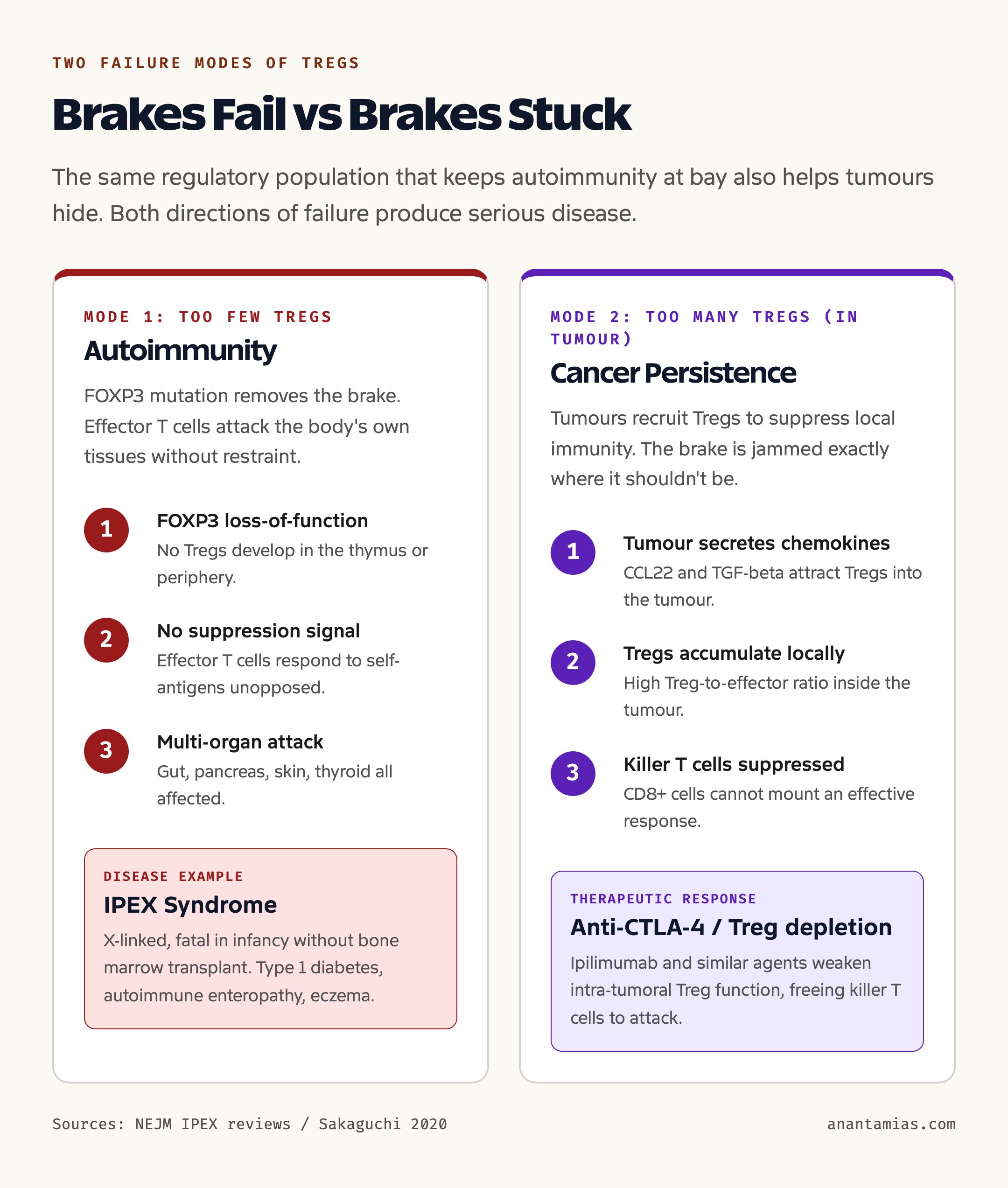
Without functional Tregs, the immune system attacks everything that moves, including the body itself. The most dramatic illustration is IPEX syndrome. Boys born with a loss-of-function mutation in FOXP3 develop, in the first months of life, severe autoimmune enteropathy that destroys the gut lining, type 1 diabetes from autoimmune destruction of pancreatic beta cells, eczema, thyroiditis and other organ-specific autoimmunity. Without bone marrow transplantation, most affected infants die in the first two years. The single-gene nature of IPEX, and the severity of the resulting disease, was the clinical evidence that finally proved Tregs were not optional.
The same biology underlies more common autoimmune diseases at a less catastrophic scale. Type 1 diabetes, multiple sclerosis, rheumatoid arthritis, lupus and inflammatory bowel disease are all associated with quantitative or qualitative defects in Treg function. Therapies that expand or stabilise Tregs, including low-dose IL-2 and adoptive Treg transfer, are in clinical trials for several of these conditions. Transplantation medicine is similarly invested. A reliable way to expand the recipient’s own Tregs against a transplanted organ would, in principle, allow the immune system to accept the graft without lifelong general immunosuppression.
The flip side of the story is cancer. A solid tumour is a hostile environment for the immune system, and tumours have learned to recruit Tregs to make themselves more hostile still. Tregs accumulate in tumour tissue, suppress the killer T cells that would otherwise destroy cancer cells, and create a pocket of local immune privilege. Tumour-infiltrating Tregs are associated with worse prognosis in many cancers. Cancer immunotherapy now includes strategies to deplete or weaken Tregs in the tumour microenvironment, including antibodies against CTLA-4 and CCR4, while leaving Tregs in healthy tissue alone. Getting that selectivity right is one of the central problems in oncology today.
Detailed Analysis: From Lab to Clinic
The path from Sakaguchi’s 1995 paper to a clinical product takes longer than most public discussion of medicine assumes. For Tregs, it has unfolded along three tracks. The first is diagnostic. Identifying Tregs reliably is now routine in research and in some clinical trials, using a combination of CD4, CD25, low CD127 and intracellular FOXP3 staining, with epigenetic confirmation by Treg-specific demethylated region analysis. This has made it possible to ask whether a particular disease has too few or too many Tregs and whether a candidate therapy moves the dial.
The second track is cellular therapy. Adoptive transfer of expanded Tregs is being tested in autoimmune disease, in graft-versus-host disease after stem cell transplant, and in solid organ transplantation. Several early-phase trials have shown safety and biological signal, though demonstrating durable clinical benefit in larger trials remains the challenge. Engineered Tregs, including chimeric antigen receptor Tregs that target the regulatory cell to a specific tissue or graft, are an active frontier. A successful CAR-Treg, in principle, could re-tolerise an autoimmune attack to a single organ without globally weakening the immune system, see our coverage of related work in stem cell research.
The third track is small-molecule and biologic modulation of Treg function in cancer. Anti-CTLA-4 antibodies, of which ipilimumab is the prototype, work in part by reducing the suppressive effect of intra-tumoral Tregs. Newer approaches aim for deeper Treg depletion in the tumour, with sparing of peripheral Tregs that maintain tolerance elsewhere. The therapeutic window is narrow because the same population that keeps cancer alive also keeps autoimmunity at bay, and aggressive systemic Treg depletion produces immune-related adverse events that mirror IPEX in their tissue distribution.
Comparative Snapshot: Effector T Cells, Helper T Cells, Tregs
| Feature | Effector CD8 T Cell | Helper CD4 T Cell | Regulatory T Cell |
|---|---|---|---|
| Lineage role | Kill infected or malignant cells | Coordinate immune response | Suppress immune response |
| Surface markers | CD3+ CD8+ | CD3+ CD4+ | CD3+ CD4+ CD25+ CD127-low |
| Master transcription factor | T-bet, others | T-bet, GATA3, RORgt, depending on subset | FOXP3 |
| Key cytokines produced | IFN-gamma, granzymes, perforin | IL-2, IL-4, IL-17, others | IL-10, TGF-beta |
| Activation outcome | Cytotoxic killing | Amplification of immune response | Brake on immune response |
| Frequency among CD4+ T cells | Not applicable | Majority | Roughly 5 to 10 percent |
| Disease association | Loss leads to chronic infection, cancer | Imbalance leads to inflammation | Loss leads to autoimmunity, gain shields tumours |
Challenges and Open Questions
The first challenge is heterogeneity. Tregs are not a single homogeneous population. They include thymic and induced subsets, tissue-resident populations adapted to specific organs, and various activation states. Designing therapies that target one subset without disturbing another is an open problem.
The second is stability. FOXP3 expression can be unstable in some contexts, particularly in inflamed tissue, and a Treg can convert into an effector-like cell that retains memory of the antigen but has lost its suppressive programme. Such ex-Tregs can drive rather than suppress disease. Locking in the regulatory phenotype, including via the epigenetic demethylated region signature, is a goal of cellular therapy.
The third is selectivity in cancer. Tumour-infiltrating Tregs and peripheral Tregs share most of their molecular machinery. Antibodies and small molecules that hit one population usually hit the other. The hunt for tumour-selective Treg targets is ongoing, with candidates including specific chemokine receptors, metabolic vulnerabilities and checkpoint molecules expressed preferentially on intra-tumoral Tregs.
The fourth is access and cost. Cellular therapies are expensive and require specialised manufacturing infrastructure. For India, the question is whether Treg therapies, when they mature, can be produced and delivered within a public health system that is also still expanding access to basic care. Indigenous capability in cell manufacturing, of the kind being built around CAR-T cell therapy in Mumbai and Bengaluru, will determine whether Treg therapy follows the same domestic path or remains an import.
Prelims Pointers
- FOXP3 stands for Forkhead Box P3 and is located on the X chromosome.
- FOXP3 is the master transcription factor for regulatory T cells.
- Regulatory T cells are CD4+ CD25+ FOXP3+ and make up roughly 5 to 10 percent of CD4+ T cells.
- Tregs were discovered by Shimon Sakaguchi in 1995.
- IPEX syndrome is the X-linked disease caused by loss of FOXP3 function.
- The scurfy mouse model led to the identification of FOXP3 as the gene mutated in IPEX.
- Tregs maintain peripheral immune tolerance, distinct from central tolerance in the thymus.
- The 2025 Nobel Prize in Physiology or Medicine, as per latest available data, was awarded for the discovery of peripheral immune tolerance and regulatory T cells.
- The Treg-specific demethylated region is the epigenetic signature of stable Tregs.
- Anti-CTLA-4 cancer immunotherapy works in part by reducing intra-tumoral Treg function.
Mains Practice Questions
- Discuss the role of regulatory T cells in maintaining peripheral immune tolerance. How does the discovery of FOXP3 explain both autoimmune disease and the persistence of solid tumours? (250 words, GS-III)
- Explain the concept of peripheral immune tolerance. How does it differ from central tolerance, and what are the consequences of its failure? (150 words, GS-III)
- “Cancer immunotherapy is, in part, the science of taking the brakes off the immune system.” Examine this statement with reference to the role of regulatory T cells and checkpoint inhibitors. (250 words, GS-III)
- India’s biotechnology ecosystem must invest in cellular therapy manufacturing if it is to participate in the next wave of immunological medicine. Comment. (150 words, GS-III)
Way Forward
The next decade of Treg biology will be shaped by three trends. The first is precision targeting. The crude tools of yesterday, broad immunosuppressants and broad immunostimulants, are giving way to therapies that act on specific cell populations in specific tissues. Engineered Tregs that home to the pancreas to halt type 1 diabetes, or to a transplanted kidney to prevent rejection, are no longer science fiction. They are in early-phase clinical trials.
The second is the integration of Treg biology with the broader story of immune regulation. Other regulatory cell types, including regulatory B cells and tolerogenic dendritic cells, work alongside Tregs in tissue-specific networks. Understanding how these populations cooperate, and how they can be co-modulated, is a frontier. So is the connection to the microbiome, which shapes the induced Treg pool through metabolites such as short-chain fatty acids.
The third is the public-health translation in countries like India. The diseases that Treg therapy may eventually address, autoimmune diabetes, lupus, inflammatory bowel disease, transplant rejection and certain cancers, are already significant burdens in India and growing. A domestic capability in Treg-based therapy, built on the scaffolding now being assembled for CAR-T cells and other cell-based therapies, will determine whether Indian patients can access these treatments at scale.
For students, the key takeaway is that FOXP3 and Tregs sit at the conceptual centre of modern immunology. The same biology explains why the immune system does not normally attack the body, why some unfortunate infants face fatal autoimmunity, why cancer is harder to treat than infection, and why a single transcription factor can be both a target and a tool of medicine.
Frequently Asked Questions
What is the FOXP3 gene and where is it located?
FOXP3, or Forkhead Box P3, is a gene located on the short arm of the X chromosome at Xp11.23. It encodes a transcription factor, which is a protein that controls the expression of other genes. FOXP3 is the master regulator of the regulatory T cell lineage and is essential for the development and function of these cells.
What are regulatory T cells (Tregs)?
Regulatory T cells are a specialised subset of CD4+ T cells that suppress immune responses. They make up roughly 5 to 10 percent of CD4+ T cells in healthy adults. Their primary role is to prevent the immune system from attacking the body’s own tissues, a function called peripheral immune tolerance. They are identified by the markers CD4, CD25, low CD127, and intracellular FOXP3.
What is IPEX syndrome?
IPEX, or Immunodysregulation Polyendocrinopathy Enteropathy X-linked, is a rare and severe autoimmune disease caused by loss-of-function mutations in the FOXP3 gene. Affected boys develop severe autoimmune enteropathy, type 1 diabetes, eczema and other autoimmune conditions in the first months of life. Without bone marrow transplantation, most do not survive past the first few years.
Who discovered regulatory T cells?
Shimon Sakaguchi at Nagoya University reported in 1995 that a population of CD4+ CD25+ T cells could prevent autoimmune disease in mice. This is generally credited as the foundational discovery of regulatory T cells. The link to FOXP3 was established later, in part through work by Mary Brunkow and Fred Ramsdell on the scurfy mouse model.
What does the 2025 Nobel Prize in Physiology or Medicine recognise?
As per latest available data, the 2025 Nobel Prize in Physiology or Medicine recognises the discovery of peripheral immune tolerance and the role of regulatory T cells, with the prize awarded to Mary Brunkow, Fred Ramsdell and Shimon Sakaguchi for the chain of work that established the existence, identity and molecular basis of this regulatory cell population.
How do Tregs prevent autoimmune disease?
Tregs suppress effector T cells and other immune cells that would otherwise attack the body’s own tissues. They do this through several mechanisms, including the secretion of inhibitory cytokines such as IL-10 and TGF-beta, the consumption of IL-2 that other T cells need, the expression of inhibitory surface molecules such as CTLA-4, and direct cell-to-cell contact that switches off effector responses.
Why do tumours attract Tregs?
Tumours create a microenvironment that favours Treg recruitment, partly through the release of chemokines and partly through the secretion of TGF-beta and other tolerogenic signals. Tregs that accumulate in tumour tissue suppress the killer T cells that would otherwise destroy cancer cells. High densities of tumour-infiltrating Tregs are associated with worse prognosis in many cancers and are a target of cancer immunotherapy.
Are Treg therapies available for patients today?
Treg-based therapies are still largely investigational. Early-phase clinical trials are testing adoptive transfer of expanded Tregs in conditions such as graft-versus-host disease, type 1 diabetes and solid organ transplantation. Engineered Tregs, including CAR-Tregs, are at an even earlier stage. Standard-of-care use of Treg-based cellular therapy is not yet established for most conditions.
How are Tregs identified in research and clinical labs?
Tregs are identified by a combination of surface markers and intracellular staining. The standard panel includes CD4 and CD25 on the surface, low CD127, and intracellular FOXP3. For more rigorous identification, the methylation status of the Treg-specific demethylated region in the FOXP3 locus is analysed, since stable Tregs show full demethylation of this region.
Why is FOXP3 on the X chromosome significant?
The X-linked location of FOXP3 explains why IPEX syndrome affects boys far more severely than girls. Boys have only one X chromosome and therefore one copy of FOXP3. A loss-of-function mutation produces full disease. Girls have two X chromosomes, so a single mutated copy is usually compensated for by the other. This pattern was a clinical clue that helped map the IPEX gene.
Tell Google you want more of this.
Add Anantam IAS as a preferred sourceOne tap, and this site shows up more often in your own Top Stories, AI Overviews and AI Mode. Remove it any time.