
Why in News?
Researchers at the Indian Institute of Science, working with clinicians from MS Ramaiah Medical College and Hospitals, reported that low-frequency ultrasound could selectively trigger death in patient-derived oral-cancer cells under laboratory conditions.
The Indian Express discussed the work as a possible future treatment route, but the paper in Materials Today Bio is an ex vivo and in vitro proof-of-concept. It did not administer ultrasound to patients, demonstrate tumour shrinkage in a living organism, or establish a clinically approved oral-cancer therapy.
- The study used cells isolated from surgically removed oral tumour samples collected between January 2023 and December 2025, including cancers from different oral sites and stages.
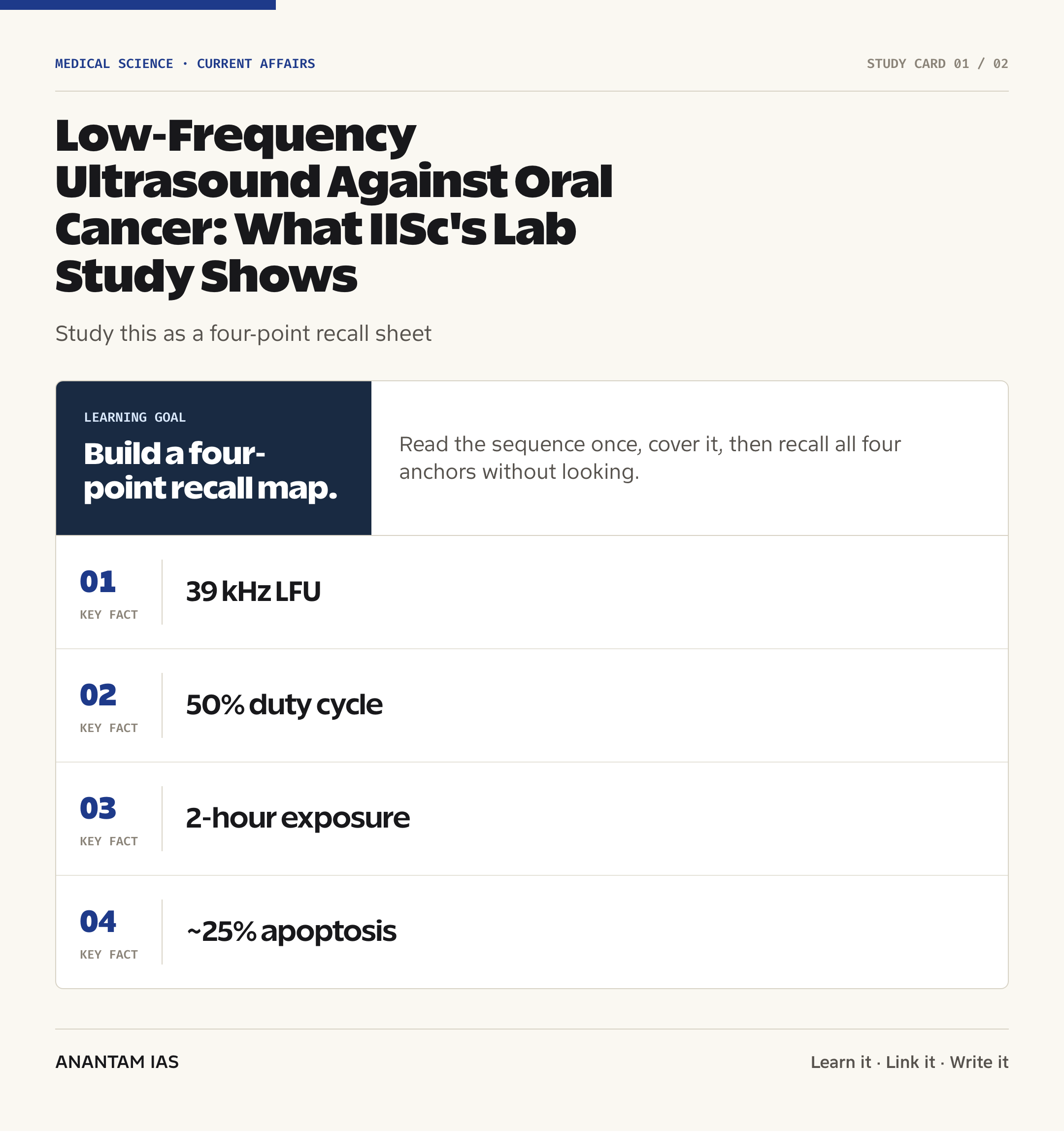
- A custom device delivered ultrasound at 39 kHz, generally at a 50% duty cycle for two hours, while experiments varied acoustic pressure and exposure time.
- At 50 kPa and 75 kPa, the paper reported about 25% and 28% apoptosis, respectively, after two hours; normal control cells showed about 5% apoptosis.
- The proposed vulnerability involves reduced Tropomyosin 2.1, or Tpm2.1, a cytoskeletal mechanosensory protein that helps cells withstand mechanical forces.
- In a simplified 3D co-culture model, ultrasound also weakened interactions between cancer cells and cancer-associated fibroblasts that can form a stromal barrier around a tumour core.
- The authors call for testing in organoids, complex tumour models and animals before any claim about safety or effectiveness in patients can be made.
The development matters in the context of:
- Oral cancer is a major Indian public-health concern associated strongly with smoked and smokeless tobacco, areca nut and alcohol; prevention and early detection remain essential regardless of this experimental result.
- Current care may combine surgery, radiotherapy and systemic therapy, depending on site, stage and patient factors, but treatment can affect speech, swallowing, appearance and quality of life.
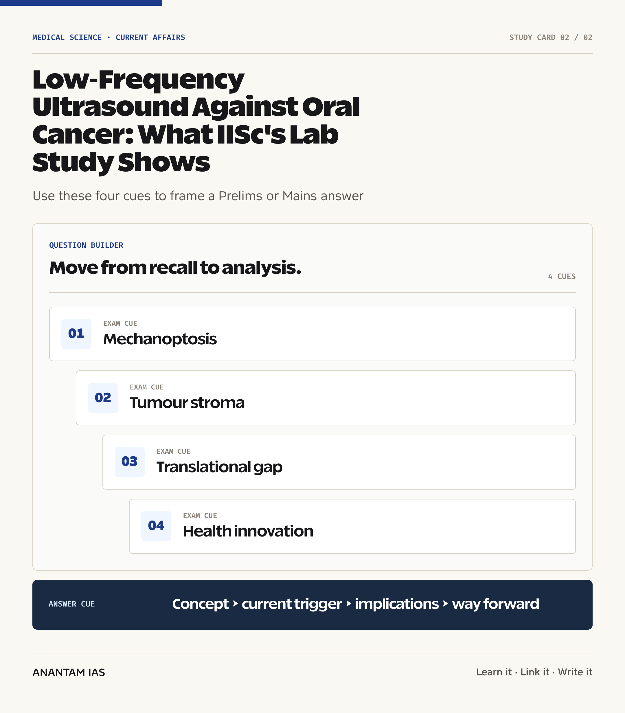
- The study is relevant to translational research: promising cellular selectivity must survive tests of tissue complexity, dosage, device delivery, toxicity, recurrence and long-term outcomes.
- It also illustrates mechanobiology, which studies how physical forces and the mechanical properties of cells influence biological behaviour and disease.
UPSC Relevance
Prelims Relevance
- Ultrasound consists of sound waves above the upper frequency limit of human hearing; frequency, intensity, duty cycle and exposure time shape its biological effects.
- Low-frequency ultrasound in this study was used as mechanical stimulation, not as a diagnostic image and not as high-intensity focused ultrasound thermal ablation.
- Apoptosis is regulated cell death; the paper uses the term mechanoptosis for apoptosis induced by mechanical forces in mechanically vulnerable cells.
- Tropomyosin 2.1 is associated with cytoskeletal rigidity sensing and actin-myosin function; reduced levels correlated with greater ultrasound sensitivity.
- MicroRNA-21 is a small non-coding RNA. The study found higher miR-21 expression in oral-cancer cells and linked it to reduced Tpm2.1 expression.
- Focal adhesions connect a cell’s cytoskeleton to the extracellular matrix and participate in mechanotransduction, the conversion of a physical cue into a biochemical response.
- Myosin IIA supports actomyosin contractility used in cell movement and invasion; its fibre organisation was disrupted after ultrasound exposure in the experiments.
- Cancer-associated fibroblasts, or CAFs, are stromal cells that can deposit extracellular matrix, influence tumour behaviour and obstruct drug or immune-cell access.
- A 3D co-culture grows more than one cell type in a spatial model; it is more informative than a simple monolayer but doesn’t reproduce a complete living tumour.
- Ex vivo research studies cells or tissues removed from an organism, while in vivo research studies effects within a living organism.
Mains Relevance
GS Paper 3
- Explain how mechanobiology and bioengineering can identify physical vulnerabilities in cancer cells and create new routes for targeted therapy.
- Assess the translational path from patient-derived cells to organoids, animal studies, device standardisation and phased clinical trials.
- Examine the role of collaboration among research institutes, hospitals and engineers in converting laboratory science into affordable health technology.
GS Paper 2
- Connect oral-cancer innovation with prevention, screening, referral and equitable cancer care rather than treating a future device as a substitute for public health.
- Discuss safeguards for evidence-based communication so that an early laboratory result doesn’t become medical misinformation or encourage patients to delay established care.
- Evaluate how public research funding can support validation while preserving patient safety, ethics and access in health-technology development.
Essay
- The distance between a scientific possibility and a public-health solution
- Innovation is credible when curiosity is matched by evidence and restraint
- Technology can improve care only when prevention and access advance with it
Background and Context
What the experiment actually tested
The work tested how cultured oral-cancer cells respond to controlled acoustic mechanical forces outside the human body.
- The team isolated primary cells from fresh, surgically removed oral tumour samples and cultured them at IISc; samples represented oral sites such as the buccal mucosa, gingivobuccal mucosa and alveolus.
- The paper describes samples from patients across cancer stages I-IV. Individual figures often draw on cells from only two or three patient samples, which matters when judging the breadth of each result.
- The custom ultrasound apparatus used a Langevin piezoelectric transducer. Cell-culture dishes were partly submerged in degassed water, and pressure at the dish was measured with a hydrophone.
- The standard setting was 39 kHz, 50% duty cycle and two hours, unless a particular experiment varied time, pressure or duty cycle. These are laboratory parameters, not a recommended dose for a patient.
- At 50 kPa and 75 kPa, annexin-V testing showed about 25% and 28% apoptosis in oral-cancer cells after two hours. This is evidence of partial cell death, not complete destruction of a tumour.
- The culture medium stayed near 25°C, with a reported change of about ±1.5°C. That observation supports a mainly mechanical rather than heat-driven effect in this setup.
How mechanical selectivity may work
The proposed selectivity rests on differences in how malignant and normal cells sense and resist physical stress.
- Cells detect force through the cytoskeleton, focal adhesions and mechanosensitive channels. Healthy cells with intact force-sensing machinery can adapt to moderate stress more effectively.
- Patient-derived oral-cancer cells showed lower levels of Tpm2.1 than normal controls. Tpm2.1 helps regulate actin-myosin interactions and rigidity sensing.
- The study found roughly a four-fold rise in miR-21 expression in oral-cancer cells compared with normal cells. The authors connect this oncogenic microRNA with suppression of Tpm2.1.
- Knocking down Tpm2.1 in normal MCF 10A cells increased their vulnerability to ultrasound, strengthening the proposed link between Tpm2.1 depletion and mechanoptosis.
- The authors discuss calcium entry through mechanosensitive channels and calpain activity as a plausible route to focal-adhesion breakdown, but the precise pathway needs deeper validation in oral tumours.
- Normal controls showed about 5% apoptosis, while cancer cells showed about 25-28% under the cited pressure settings. ‘Selective’ doesn’t mean that every cancer cell died or that every normal cell was unaffected.
Effects beyond cell survival
The researchers also examined cell structures and behaviours associated with movement and invasion.
- Ultrasound reduced the number and area of mature focal adhesions in oral-cancer cells and produced a shrunken morphology, while normal cells showed a different adaptive response.
- It disassembled myosin IIA fibres, weakening actomyosin contractility that cancer cells use to reshape themselves and move through surrounding material.
- In collagen-based laboratory assays, treated cancer cells showed reduced migration, gel contraction and invasion. These assays model components of metastatic behaviour but don’t prove reduced metastasis in a person.
- This distinction matters: an assay can show a lower capacity to migrate through a model matrix, while clinical metastasis also depends on blood vessels, immune interactions, tissue barriers and many molecular signals.
- The work fits a wider field of targeted biomedical innovation that includes nanomedicine and targeted drug delivery, but ultrasound here acts through physical stimulation rather than carrying a drug.
- It also differs from CAR T-cell therapy, which engineers immune cells. The comparison shows that ‘precision treatment’ can refer to very different mechanisms and evidence pathways.
What the 3D co-culture adds
A second part of the study modelled interactions between tumour aggregates and cancer-associated fibroblasts on an elastomeric platform.
- The platform placed 300-500 micrometre tumour aggregates with patient-derived CAFs inside small PDMS wells, allowing time-lapse observation of encapsulation, infiltration and the tumour-stroma interface.
- CAFs can produce extracellular matrix and form a capsule-like stromal barrier. Such a barrier may limit penetration by drugs and immune cells and can influence tumour organisation.
- After one hour of ultrasound and eight hours of imaging, the model showed an approximately 80% reduction in CAF capsule intensity in the encapsulation region.
- CAF invasion speed declined, and the distance travelled by infiltrating CAFs was about half that of untreated controls. CAF intensity at the tumour interface fell by roughly 33%.
- These numbers describe fluorescent intensity and movement in a simplified laboratory platform; they aren’t measures of drug delivery, immune infiltration or treatment response in a patient.
- The model isn’t an organoid. The authors describe it as a reductionist co-culture containing cancer cells and CAFs, without immune cells, endothelial cells or the full architecture and physiology of an oral tumour.
Why the normal-cell comparison needs caution
Control choice is central to the claim that the intervention discriminates between malignant and healthy tissue.
- The experiments used patient-derived normal oral epithelial cells where available, but several analyses also used MCF 10A, a non-tumourigenic breast epithelial cell line, as the normal comparator.
- The paper itself identifies inadequate access to matched healthy oral mucosal cells as a limitation and recommends adjacent normal oral tissue or tissue collected during suitable dental procedures.
- A living oral cavity contains nerves, blood vessels, bone, salivary tissue, immune cells and microbiota. A safe acoustic window must protect all relevant tissues, not just cultured epithelial controls.
- Differences in tumour depth, geometry, stiffness and location could alter acoustic pressure distribution. A setting that works at the bottom of a culture dish may not reproduce its effect through real tissue.
- Longer-term questions include inflammation, wound healing, recurrence, effects on residual normal tissue and whether mechanical exposure can alter cell dispersal. None was resolved by this study.
- So the responsible conclusion is selective activity in tested models, followed by a need for independent replication and preclinical safety studies.
From proof-of-concept to possible therapy
Translation would require a staged evidence programme rather than direct adoption of the laboratory apparatus.
- Researchers first need reproducible results across a larger and clinically diverse bank of patient-derived cells and matched controls, with transparent dose-response and statistical reporting.
- More realistic systems should include organoids, immune components, vasculature and extracellular matrix, followed by animal models capable of showing tissue-level safety and tumour response.
- Engineers must establish dosimetry, beam geometry, coupling, treatment duration and real-time monitoring for an intraoral delivery device that can reach irregular lesions without unsafe hotspots.
- Combination studies can test whether stromal disruption improves penetration of an established drug, but improved fluorescence patterns alone don’t prove a better therapeutic outcome.
- Human trials would need phased evaluation of safety, dose, efficacy and comparison with standard care. Until those steps succeed, clinicians shouldn’t replace surgery, radiation or systemic therapy with this method.
- India’s wider biotechnology ecosystem can help link hospital samples, bioengineering, clinical oncology, manufacturing and regulation, while affordability must be designed from the start.
Public-health significance for India
A future targeted treatment would address only one part of India’s oral-cancer challenge.
- Reducing exposure to smokeless tobacco, smoking, areca nut and harmful alcohol use remains the strongest population-level route to lowering preventable disease.
- Persistent mouth ulcers, red or white patches, unexplained bleeding, a lump or difficulty swallowing require timely professional evaluation; experimental headlines shouldn’t encourage self-treatment or delay.
- Primary care and dental systems need stronger risk counselling, visual examination, referral and pathology access, especially for people with prolonged tobacco or areca-nut exposure.
- Rehabilitation after treatment, including nutrition, speech, swallowing, dental and psychosocial support, is part of quality cancer care, not an optional extra.
- If ultrasound mechanostimulation reaches clinical use, its value should be judged against standard care through survival, recurrence, function, adverse effects, cost and quality-of-life outcomes.
- The study’s real present value is scientific: it identifies a testable biomechanical vulnerability and a route for further research, not a treatment available to patients today.
Way Forward
Strengthen biological validation
- Repeat the findings across more matched oral-cancer and healthy oral samples, stages, anatomical sites and relevant molecular subtypes.
- Test the Tpm2.1-miR-21 mechanism using complementary genetic and pharmacological methods, and publish the full range of responders and non-responders.
Move through realistic preclinical models
- Progress from the simplified platform to organoids and multicellular models containing stroma, immune cells, vasculature and clinically realistic extracellular matrix.
- Use animal studies to assess tissue injury, inflammation, cell dispersal, tumour control, recurrence and interaction with surgery, radiation or medicines.
Standardise the technology
- Define reproducible frequency, pressure, duty cycle and exposure ranges and map how tissue depth and shape change the delivered acoustic dose.
- Develop an ergonomic intraoral applicator with monitoring and shut-off safeguards before considering carefully governed early-phase human testing.
Communicate without hype
- Label the result consistently as a laboratory proof-of-concept and separate observed findings from hypotheses about drug penetration or future clinical benefit.
- Pair innovation reporting with clear advice that patients should continue evidence-based evaluation and treatment under qualified oncology teams.
Keep prevention and access central
- Sustain tobacco and areca-nut control, early detection, pathology capacity, referral networks and rehabilitation while experimental therapies are developed.
- Build affordability, Indian manufacturing and equitable trial recruitment into the translation plan so a successful device doesn’t widen access gaps.
Conclusion
The IISc low-frequency ultrasound study offers a credible mechanobiology hypothesis: oral-cancer cells with weakened force-sensing machinery may be more vulnerable than normal cells to controlled mechanical stimulation. Patient-derived cells, explicit acoustic parameters and a 3D cancer-cell/CAF platform make the proof-of-concept worth following.
But the evidence remains outside the human body and is based on simplified models. It neither cures oral cancer nor establishes that ultrasound is safe and effective in patients. The next scientific test is whether selectivity survives organoid, animal and device-level validation without harming the complex tissues of the mouth.
For public policy, the lesson is two-track: support rigorous translational research, and keep investing in prevention, early diagnosis and accessible standard treatment. Promising science earns trust when its limits are explained as carefully as its possibilities.
UPSC Practice Questions
Prelims MCQ 1
With reference to the IISc study on low-frequency ultrasound and oral-cancer cells, consider the following statements:
- It tested cultured patient-derived cancer cells and a simplified 3D co-culture platform.
- It linked reduced Tropomyosin 2.1 with greater sensitivity to mechanical stimulation.
- It established superior survival outcomes in a randomised clinical trial.
How many of the above statements are correct?
(a) Only one (b) Only two (c) All three (d) None
Answer: (b) Only two
Explanation:
Statements 1 and 2 are correct. The work was an ex vivo and in vitro proof-of-concept. It included no treatment of patients and no randomised clinical trial, so it couldn’t establish survival benefit.
Prelims MCQ 2
Which one of the following best describes mechanoptosis in the study?
(a) Thermal burning of tissue by high-intensity focused ultrasound (b) Imaging a tumour through reflected high-frequency sound waves (c) Apoptosis triggered by mechanical forces in mechanically vulnerable cells (d) Drug release from a capsule dissolved by body heat
Answer: (c) Apoptosis triggered by mechanical forces in mechanically vulnerable cells
Explanation:
Mechanoptosis refers to regulated cell death induced by mechanical stimulation. The IISc team sought a non-thermal effect and associated cancer-cell vulnerability with reduced Tpm2.1-mediated mechanosensing.
UPSC Mains Questions
- The IISc study on low-frequency ultrasound and oral-cancer cells shows both the value and the limits of patient-derived laboratory models. Explain its proposed mechanobiological mechanism and evaluate the evidence still required before such a method can enter clinical oncology. (15 marks, 250 words)
- Biomedical innovation should be assessed not only by novelty but also by the strength of its translational pathway. Discuss with reference to model complexity, dosimetry, safety, clinical trials, regulatory oversight and affordability in ultrasound-based cancer research. (15 marks, 250 words)
- A promising cancer-treatment headline must not displace prevention and established care. Examine how India can support high-risk health research while strengthening tobacco control, early oral-cancer detection, referral, rehabilitation and responsible public communication. (15 marks, 250 words)
Sources: Indian Institute of Science and The Indian Express Explained.
Frequently Asked Questions
Has IISc developed an oral-cancer cure?
No. IISc researchers demonstrated selective effects on cultured patient-derived oral-cancer cells and a simplified 3D co-culture model. They didn’t treat patients or show tumour control in an animal. The method needs organoid studies, animal validation, device development, safety testing and clinical trials before it could become a treatment.
What is low-frequency ultrasound mechanostimulation?
It is the use of low-frequency acoustic waves to apply controlled mechanical forces to cells or tissues. In this study, the researchers used 39 kHz ultrasound and sought a mainly mechanical, non-thermal effect. That laboratory setup is different from diagnostic imaging and from high-intensity focused ultrasound used for thermal ablation.
Why were cancer cells more vulnerable?
The study links vulnerability to reduced Tropomyosin 2.1, a cytoskeletal protein involved in sensing rigidity and resisting mechanical stress. Higher miR-21 expression may suppress this protein. When exposed to ultrasound, mechanically vulnerable cells showed focal-adhesion disruption, loss of myosin organisation and higher apoptosis than normal controls.
Did ultrasound kill every cancer cell?
No. After two hours, the paper reported about 25% apoptosis at 50 kPa and 28% at 75 kPa, compared with roughly 5% in normal controls. These are results from cell cultures under defined conditions. They don’t mean that an entire tumour would disappear or that all normal tissue would be spared.
What did the 3D model demonstrate?
The model brought patient-derived tumour aggregates together with cancer-associated fibroblasts. Ultrasound reduced fibroblast encapsulation, infiltration and accumulation at the tumour interface. But it was a reductionist PDMS co-culture, not an organoid or a living tumour, and it lacked immune and endothelial cells.
Should patients seek this ultrasound treatment now?
No. This experimental method isn’t an established clinical treatment. Anyone with a suspicious oral lesion or a cancer diagnosis should seek prompt evaluation from qualified dental, surgical and oncology professionals and follow evidence-based care. Prevention, early detection and timely treatment remain the practical priorities today.
Tell Google you want more of this.
Add Anantam IAS as a preferred sourceOne tap, and this site shows up more often in your own Top Stories, AI Overviews and AI Mode. Remove it any time.