
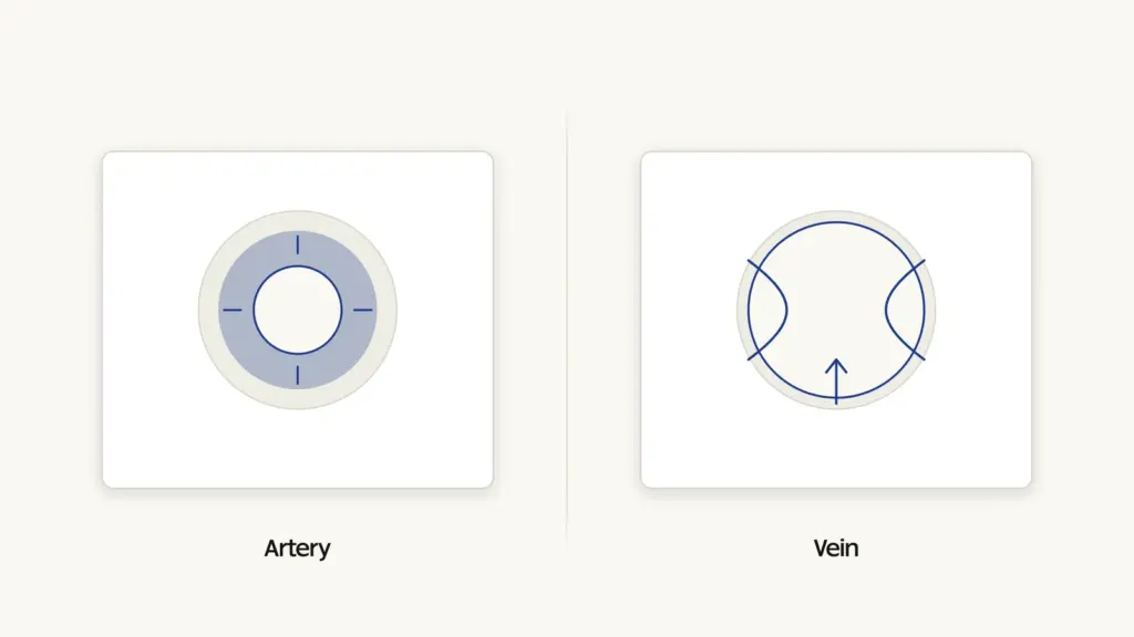
The difference between arteries and veins is defined by direction, not by the kind of blood they carry. Arteries carry blood away from the heart; veins carry blood back towards it. Everything else about them — the thickness of the wall, the width of the lumen, the presence of valves, the pressure inside — follows from that one functional difference.
The common belief that arteries carry oxygenated blood and veins carry deoxygenated blood is true of most vessels but is not the definition, and it fails at exactly the place where the circulation matters most. The pulmonary artery carries deoxygenated blood, and the pulmonary veins carry oxygenated blood. Direction is the rule; oxygenation is a pattern with exceptions.
How blood moves through the body
The human circulatory system is a closed double circulation. Blood passes through the heart twice in one complete circuit: once through the right side on its way to the lungs, and once through the left side on its way to the rest of the body.
In the systemic circuit, oxygenated blood leaves the left ventricle through the aorta, the largest artery in the body. The aorta branches into smaller arteries, then into arterioles, and finally into capillaries — vessels so narrow that red blood cells pass through in single file, with walls one cell thick. Exchange happens only at the capillaries: oxygen and nutrients diffuse out to the tissues, carbon dioxide and waste diffuse in. Capillaries then merge into venules, venules into veins, and the veins deliver blood back to the right atrium through the superior and inferior vena cava.
In the pulmonary circuit, deoxygenated blood leaves the right ventricle through the pulmonary artery, is oxygenated in the alveolar capillaries of the lungs, and returns to the left atrium through the pulmonary veins. The full pathway, along with how pressure is generated and measured, is covered in detail in this guide to the human circulatory system and blood pressure.
Arteries and veins: comparison table
| Basis | Arteries | Veins |
|---|---|---|
| Direction of flow | Carry blood away from the heart | Carry blood towards the heart |
| Type of blood carried | Oxygenated, except the pulmonary artery and the umbilical arteries | Deoxygenated, except the pulmonary veins and the umbilical vein |
| Wall thickness | Thick and strong, with a well-developed muscular middle layer | Thin, with much less muscle and elastic tissue |
| Elasticity | Highly elastic; the wall stretches and recoils with each heartbeat | Poorly elastic; the wall is largely fibrous |
| Lumen | Narrow lumen relative to wall thickness | Wide lumen relative to wall thickness |
| Valves | Absent, except the semilunar valves where the aorta and pulmonary artery leave the heart | Present along the length, especially in the limbs, preventing backflow |
| Blood pressure | High; measured at about 120/80 mm Hg in a healthy adult | Low, falling to near zero at the entry to the heart |
| Nature of flow | Rapid and pulsatile, in spurts matching the heartbeat | Slow and steady, without a pulse |
| Position in the body | Mostly deep-seated, protected by muscle and bone | Many are superficial and visible under the skin |
| Behaviour when empty | Remain open because of the thick elastic wall | Collapse when not filled |
| Effect of injury | Blood escapes in spurts under pressure; bleeding is rapid and dangerous | Blood flows out steadily; bleeding is easier to control |
| Largest vessel | Aorta | Superior and inferior vena cava |
| Branching pattern | Divide into arterioles and then capillaries | Formed by capillaries uniting into venules and then veins |
| Blood volume held | Roughly a sixth of total blood volume at any moment | Act as a reservoir, holding the majority of circulating blood |
The structure of an artery
Every blood vessel larger than a capillary is built from three concentric layers.
The innermost layer, the tunica intima, is a single sheet of flat endothelial cells resting on a thin basement membrane. It is smooth, which minimises resistance and discourages clotting.
The middle layer, the tunica media, is where arteries and veins diverge most sharply. In an artery this layer is thick, packed with circular smooth muscle and sheets of elastin. In the aorta and the large arteries near the heart, elastin dominates: the vessel stretches when the left ventricle contracts, storing energy in its stretched wall, and recoils during relaxation, pushing blood onward. This is why blood keeps flowing between heartbeats instead of stopping and starting. Further from the heart, in arterioles, smooth muscle dominates instead, and its contraction or relaxation controls how much blood reaches a given tissue.
The outer layer, the tunica externa, is tough connective tissue that anchors the vessel and resists over-expansion.
The pulse you feel at the wrist is the elastic wall of the radial artery expanding as a pressure wave passes. Veins have no pulse, because by the time blood reaches them the pressure wave has been absorbed by the arterioles and capillaries.
The structure of a vein
A vein has the same three layers, but the proportions are reversed. The tunica media is thin, with little muscle and little elastin, because there is no high pressure to contain. The lumen is correspondingly wide, which lowers resistance to a slow-moving column of blood. An empty vein collapses into a flattened tube; an empty artery holds its shape.
The critical structural feature of veins is the valve: paired semilunar folds of the inner lining that open in the direction of the heart and close against backflow. Valves are abundant in the veins of the arms and legs, where blood must travel upward against gravity, and largely absent in the large veins of the trunk and in those of the head and neck, where gravity assists return.
Blood returns to the heart by three mechanisms working together. The residual pressure left over from the heartbeat pushes weakly. The skeletal muscle pump does most of the work: contracting leg muscles squeeze the deep veins, and the valves ensure the squeezed blood can only move heartward. The respiratory pump contributes as pressure changes in the chest during breathing draw blood inward. This is why standing still for a long time causes blood to pool in the legs, and why moving relieves it. When valves become incompetent, blood pools permanently and the vessel distends — the mechanism behind varicose veins.
One vein does not fit the standard description. The hepatic portal vein collects blood from the stomach, intestines, pancreas, and spleen and delivers it not to the heart but to the liver, where it breaks up into capillaries a second time. This portal system exists so that everything absorbed from the digestive tract — the products of the macronutrients and vitamins in a meal, along with any toxins — is processed by the liver before entering general circulation.
Where the exchange actually happens
Neither arteries nor veins exchange anything with the tissues. Their walls are far too thick. All exchange occurs in the capillaries, and the whole architecture of the circulation exists to deliver blood to them at a manageable pressure and collect it afterwards.
A capillary wall is a single layer of endothelial cells, roughly one micrometre thick, and a capillary’s diameter is about the width of a red blood cell. What arterioles do is control access: by constricting or dilating, they decide how much blood enters each capillary bed. That is how the body sends more blood to working muscles during exercise and to the digestive tract after a meal without changing the heart’s total output very much.
Because the capillary bed is where oxygen is handed over, the oxygen-carrying capacity of blood determines how much good the whole system does. Reduced haemoglobin means the same volume of blood delivers less oxygen, which is why iron-deficiency anaemia produces breathlessness and fatigue despite a normally functioning heart and normal vessels — a public health problem addressed in India through the Anaemia Mukt Bharat strategy.
Common confusions
“Veins carry blue blood.” Blood is never blue. Oxygenated blood is bright scarlet; deoxygenated blood is a darker, brownish red. Veins look bluish through the skin because of how light of different wavelengths is scattered and absorbed by tissue before it reaches your eye. Draw the blood out and it is dark red.
“Arteries always carry oxygenated blood.” The pulmonary artery, carrying blood from the right ventricle to the lungs, is deoxygenated. In the foetus, the umbilical arteries carry deoxygenated blood to the placenta and the umbilical vein returns oxygenated blood to the foetus — the reverse of the adult pattern in both cases.
“Arteries have no valves at all.” They have no valves along their length, but semilunar valves sit at the base of the aorta and the pulmonary artery, preventing blood from falling back into the ventricles when they relax. The heart sounds you hear through a stethoscope are largely these and the atrioventricular valves closing.
“Veins are weaker, so they matter less.” Veins hold most of the body’s blood at any given moment and act as a capacitance reservoir. Constricting them shifts blood into active circulation, which is one of the body’s main responses to blood loss.
“Blood pressure is the same throughout the circulation.” It falls steadily along the route. The steepest fall occurs across the arterioles, which is precisely their function: they convert a pulsatile high-pressure flow into the gentle, steady flow that fragile capillaries can survive.
“Arteries are deep and veins are superficial, always.” It is a strong tendency, not a rule. Both deep and superficial veins exist, and a few arteries run close to the surface — which is why the radial artery at the wrist and the carotid artery at the neck are the standard places to feel a pulse.
Frequently Asked Questions
Why do arteries have thicker walls than veins?
Because they receive blood directly from the contracting ventricles at high pressure. The thick muscular and elastic middle layer withstands that pressure, and its recoil between beats keeps blood moving forward.
Why do veins have valves but arteries do not?
Venous pressure is too low to guarantee forward flow, especially in the limbs where blood must move against gravity. Valves make backflow impossible, so any squeeze from surrounding muscles pushes blood only towards the heart. Arterial pressure is high enough that backflow is not a risk.
Which artery carries deoxygenated blood?
The pulmonary artery, which takes deoxygenated blood from the right ventricle to the lungs. In foetal circulation, the umbilical arteries also carry deoxygenated blood, towards the placenta.
Why is blood taken from a vein for tests and donation?
Veins are superficial, easy to locate, and hold blood at low pressure, so a puncture is easy to seal with light pressure. An arterial puncture bleeds forcefully and needs prolonged compression, so it is used only when arterial blood is specifically required.
What are capillaries and why are they necessary?
Capillaries are the smallest vessels, with walls one cell thick, forming the bridge between arterioles and venules. They are the only site where oxygen, nutrients, and wastes are exchanged with tissues; arteries and veins are purely transport channels.
What causes varicose veins?
Failure of the valves in superficial leg veins. Once valves no longer close properly, blood flows backwards and pools, stretching the vessel wall and making it visibly twisted and swollen. Prolonged standing, pregnancy, and age increase the risk.
Where is blood pressure measured, and what do the two numbers mean?
It is measured in the brachial artery of the upper arm. The first number is systolic pressure, during ventricular contraction; the second is diastolic pressure, during ventricular relaxation. A typical healthy adult reading is about 120/80 mm Hg.
Practice Questions
1. Which of the following correctly defines an artery?
a) A vessel that carries oxygenated blood
b) A vessel that carries blood away from the heart
c) A vessel that contains valves along its length
d) A vessel with a thin wall and a wide lumen
Answer: b) A vessel that carries blood away from the heart
2. The pulmonary vein is distinctive because it:
a) Carries deoxygenated blood towards the heart
b) Carries oxygenated blood towards the heart
c) Contains more valves than any other vein
d) Arises directly from the left ventricle
Answer: b) Carries oxygenated blood towards the heart
3. Valves are largely absent in which of the following?
a) Veins of the lower limb
b) Veins of the upper limb
c) Arteries along their length
d) Veins draining the calf muscles
Answer: c) Arteries along their length
4. Exchange of oxygen, nutrients, and wastes between blood and tissues takes place in the:
a) Arterioles
b) Capillaries
c) Venules
d) Large veins
Answer: b) Capillaries
5. The hepatic portal vein is unusual because it:
a) Carries oxygenated blood from the lungs
b) Contains no valves and lies superficially
c) Carries blood from the digestive organs to the liver rather than to the heart
d) Arises directly from the aorta
Answer: c) Carries blood from the digestive organs to the liver rather than to the heart
- Distinguish between arteries and veins on the basis of structure, pressure, and function, and explain how each structural feature serves the vessel’s role.
- Explain the concept of double circulation in humans and identify the two vessels whose oxygen content is exceptional to the general rule.
- “Blood pressure falls most steeply across the arterioles.” Examine why this is physiologically necessary for capillary function.
- Describe the mechanisms of venous return and discuss how their failure leads to clinical conditions such as varicose veins.
- Discuss the role of capillaries as the functional unit of the circulatory system, and explain why reduced haemoglobin impairs tissue oxygenation even when the heart and vessels are normal.
Tell Google you want more of this.
Add Anantam IAS as a preferred sourceOne tap, and this site shows up more often in your own Top Stories, AI Overviews and AI Mode. Remove it any time.